

MUSCLE
- Specialised tissue of mesodermal origin.
- About 40-50 percent of the body weight of a human adult
- Special properties à Excitability, contractility, extensibility and elasticity.
- Muscles have been classified using different criteria, namely location, appearance and nature of regulation of their activities.
- Based on their location, three types of muscles are identified : 1). Skeletal muscles 2). Visceral muscles 3). Cardiac muscles
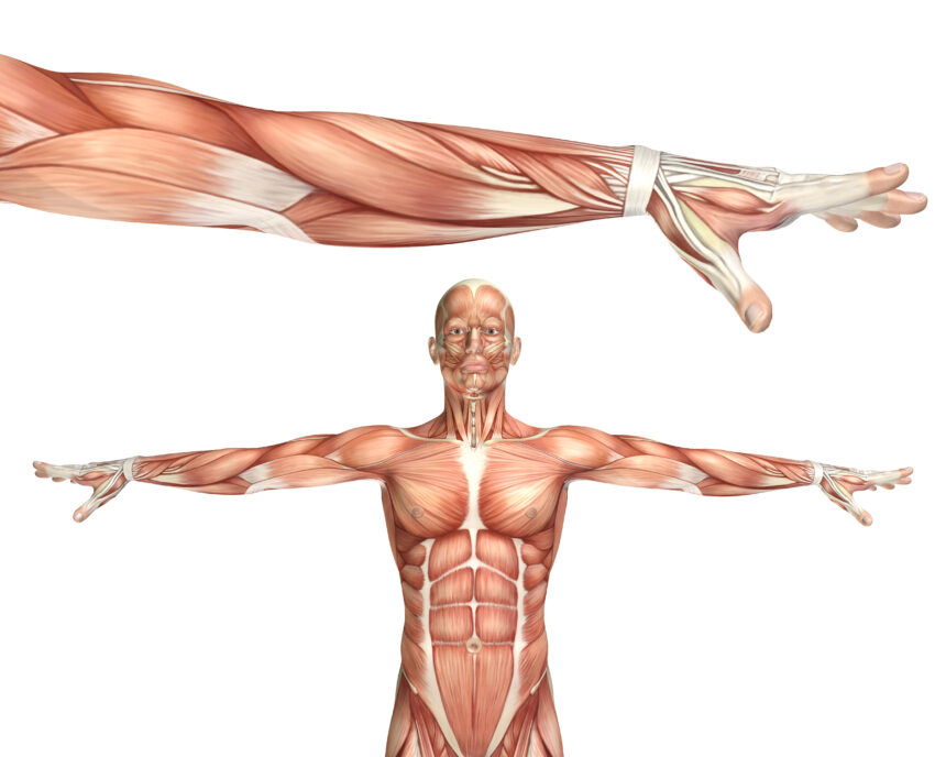
- Skeletal muscles are closely associated with the skeletal components of the body.
- They have a striped appearance under the microscope and hence are called striated muscles.
- As their activities are under the voluntary control of the nervous system, they are known as voluntary muscles too.
- They are primarily involved in locomotory actions and changes of body postures.
- Visceral muscles are located in the inner walls of hollow visceral organs of the body like the alimentary canal, reproductive tract, etc.
- They do not exhibit any striation and are smooth in appearance à Smooth muscles (nonstriated muscle).
- Not under the voluntary control of the nervous system à involuntary muscles.
- They assist, for example, in the transportation of food through the digestive tract and gametes through the genital tract.
CARDIAC MUSCLES
- Muscles of heart.
- Many cardiac muscle cells assemble in a branching pattern to form a cardiac muscle.
- Based on appearance, cardiac muscles are striated.
- Involuntary in nature as the nervous system does not control their activities directly.
SKELETAL MUSCLE
- Each organised skeletal muscle in our body is made of a number of muscle bundles or fascicles held together by a common collagenous connective tissue layer called fascia.
- Each muscle bundle contains a number of muscle fibres.
- Each muscle fibre is lined by the plasma membrane called sarcolemma enclosing the sarcoplasm.
- Muscle fibre is a syncytium as the sarcoplasm contains many nuclei.
- The endoplasmic reticulum, i.e., sarcoplasmic reticulum of the muscle fibres is the store house of calcium ions.
- A characteristic feature of the muscle fibre is the presence of a large number of parallely arranged filaments in the sarcoplasm called myofilaments or myofibrils.
- Each myofibril has alternate dark and light bands on it.
MECHANISM OF MUSCLE CONTRACTION
- Mechanism of muscle contraction is best explained by the sliding filament theory which states that contraction of a muscle fibre takes place by the sliding of the thin filaments over the thick filaments.
- Muscle contraction is initiated by a signal sent by the central nervous system (CNS) via a motor neuron.
- A motor neuron along with the muscle fibres connected to it constitute a motor unit.
- The junction between a motor neuron and the sarcolemma of the muscle fibre is called the neuromuscular junction / motor-end plate.
- A neural signal reaching this junction releases a neurotransmitter (Acetyl choline) which generates an action potential in the sarcolemma.
- Action potential spreads through the muscle fibre and causes the release of calcium ions into the sarcoplasm.
- Increase in Ca++ level leads to the binding of calcium with a subunit of troponin on actin filaments and thereby remove the masking of active sites for myosin.
- Utilising the energy from ATP hydrolysis, the myosin head now binds to the exposed active sites on actin to form a cross bridge.
- This pulls the attached actin filaments towards the centre of ‘A’ band.
- The‘Z’ line attached to these actins are also pulled inwards thereby causing a shortening of the sarcomere, i.e., contraction.
- It is clear from the above steps, that during shortening of the muscle, i.e., contraction, the ‘I’ bands get reduced, whereas the ‘A’ bands retain the length
- The myosin, releasing the ADP and P1 goes back to its relaxed state.
- A new ATP binds and the cross-bridge is broken
- The ATP is again hydrolysed by the myosin head and the cycle of cross bridge formation and breakage is repeated causing further sliding.
- The process continues till the Ca++ ions are pumped back to the sarcoplasmic cisternae resulting in the masking of actin filaments.
- This causes the return of ‘Z’ lines back to their original position, i.e., relaxation.
- The reaction time of the fibres can vary in different muscles.
- Repeated activation of the muscles can lead to the accumulation of lactic acid due to anaerobic breakdown of glycogen in them, causing fatigue.
- Muscle contains a red coloured oxygen storing pigment called myoglobin
- Myoglobin content is high in some of the muscles which gives a reddish appearance. à Red fibres.
- Red fibre muscles contain plenty of mitochondria which can utilise the large amount of oxygen stored in them for ATP production à Aerobic muscles.
- On the other hand, some of the muscles possess very less quantity of myoglobin and therefore, appear pale or whitish. à White fibres
- Number of mitochondria are also few in them, but the amount of sarcoplasmic reticulum is high. à They depend on anaerobic process for energy.